Eye Care Services

What is LASIK?

LASIK is the most common form of elective vision correction surgery in the world and can be used to treat myopia (short-sight), hyperopia (long-sight) and astigmatism.

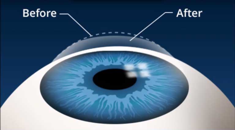
The principle of LASIK involves using a special type of laser to precisely change the shape of your cornea — the dome-shaped transparent tissue at the front of your eye — to improve vision.

Normally, images are clearly focused on the retina in the back of your eye because the light rays are bent properly to contact the retinal surface. With short-sightedness (myopia), long-sightedness (hyperopia) or astigmatism, the light is bent incorrectly and it ends up being focused elsewhere, resulting in blurred vision. Traditionally, the blurred vision is corrected by bending (refracting) light rays with glasses or contact lenses. But reshaping the cornea itself also will provide the necessary refraction.

What are the benefits of LASIK?

  • Quick, painless procedure, lasting less than 10 minutes per eye.
  • Rapid results, with patients often seeing well the next day.
  • Clinically proven treatment – Research indicates that 92-98% of patients achieve their desired vision after LASIK treatment and complete satisfaction.
  • End-to-end laser technology.
  • Bladeless surgery.
  • No stitches required.
  • Further adjustments can be made in the future to correct vision should the need arise.

LASEK is the a very popular form of elective vision correction surgery and can be used to treat myopia (short-sight), hyperopia (long-sight) and astigmatism.

The principle of LASEK involves using a special type of laser to precisely change the shape of your cornea — the dome-shaped transparent tissue at the front of your eye — to improve vision.

Normally, images are clearly focused on the retina in the back of your eye because the light rays are bent properly to contact the retinal surface. With short-sightedness (myopia), long-sightedness (hyperopia) or astigmatism, the light is bent incorrectly and it ends up being focused elsewhere, resulting in blurred vision. Traditionally, the blurred vision is corrected by bending (refracting) light rays with glasses or contact lenses. But reshaping the cornea itself also will provide the necessary refraction.

What are the benefits of LASEK?

  • Quick, painless procedure, lasting less than 5 minutes per eye.
  • Excellent results, with patients often seeing well within a few days.
  • Clinically proven treatment – Research indicates that 97% of patients achieve their desired vision after LASEK treatment and complete satisfaction.
  • No flap is created during surgery meaning it is suitable for patients with dry eyes, thin corneas or those who participate in contact sports.
  • Surgery is very comfortable with minimal touch involved.
  • No stitches required.
  • Further adjustments can be made in the future to correct vision should the need arise.

The decision to undergo private cataract surgery offers the opportunity to correct common vision problems, including short-sight, long-sight, presbyopia and astigmatism, to reduce your dependence on glasses or contact lenses permanently.

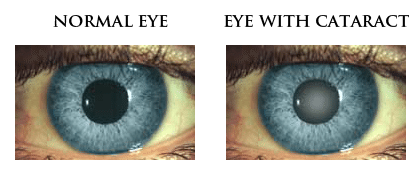
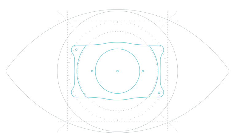
Cataract surgery involves the removal of the cloudy natural lens of the eye (the cataract) and replacing it with a carefully selected intra-ocular lens (IOL). There are several designs of intra-ocular lenses (IOLs) available, each having their own strengths in performance. The choice of lens has a major influence on the outcome of surgery and whether glasses would be required after surgery.

All our surgeons specialise in cataract surgery and have all performed several thousand cataract procedures, particularly for patients with high spectacle corrections, complex cataracts and following other types of refractive surgery.

Today, thanks to advances in technology, most patients experience little or no pain and return to their normal activities the day after surgery.

The decision to undergo cataract surgery offers a once-in-a-lifetime opportunity to insert the best lens for you. There are several designs of IOLs available, each having their own strengths in performance. The choice of lens has a major influence on the outcome of surgery and whether glasses would be required after surgery.

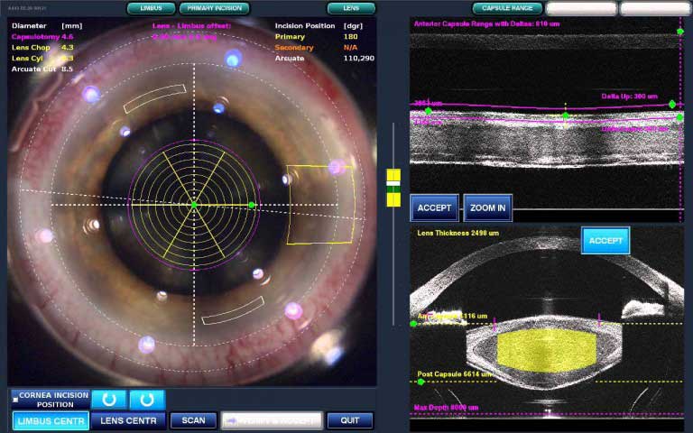
Initially used in LASIK surgery, femtosecond laser technology has evolved to offer the latest technique in performing cataract and lens replacement procedures, providing unprecedented accuracy and precision.

The Ziemer Z8 laser can now be used to complete some of the critical steps of cataract and lens replacement surgery with repeatable accuracy down to 1/1000th mm. This includes the entry incisions, capsulorhexis and softening or fragmenting the nucleus. These lasers use very short pulses (femtosecond) of energy, which create tiny gas bubbles and employ the same principles that we have been using in LASIK for some years.

For patients seeking the most advanced and accurate surgical procedure, laser cataract surgery is at the forefront and remains unsurpassed. This technology offers a reproducible technique to replace the less predictable and most technically demanding steps of conventional cataract surgery.

Refractive Lens Exchange is a particularly good choice for patients over the age of 45 years. Often at this stage, the natural lens starts to lose it’s functionality and reading glasses are required as well as distance glasses. Whilst laser may be still be a suitable option, the longevity of laser treatment is not predictable as the lens starts to change.

RLE involves the replacement of the natural lens in the eye, with a new mono-focal, multi-focal or pseudo-accommodative intra-ocular lens implant (IOL). Accurate measurements of the eye taken prior to surgery allow for a bespoke, customised intra-ocular lens (IOL) to be implanted which can correct for any degree of long or short-sightedness and astigmatism so that clear distance vision without glasses is a reality. With the choice of premium lens implants, achieving excellent reading vision without glasses may be achieved allowing for true spectacle independence.

In simple terms, this means that each patient benefits from new, personalised lenses, which provide the best and most accurate correction for their vision.

The lens exchange procedure takes between ten and fifteen minutes and is virtually painless. As with LASIK treatment, it is usually performed under local or topical anaesthetic. Patients return home the same day of their treatment and most see an immediate improvement in their vision.

Most patients will have their second eye treated within a couple of weeks and it is following this that the most dramatic results are seen.

If you are interested in finding out more about replacement lens exchange and your suitability, please contact us and we will be happy to arrange a consultation.

Lens Types

Very subtle differences in lens design mean that some perform better in certain circumstances than others and that is why accurate measurements and an in-depth consultation is needed prior to the procedure to ascertain which lenses or combination of lenses best suit you and your lifestyle.

There are several types of lens and these are namely; monofocal, multifocal (also called bifocal), accommodative, toric for the correction of astigmatism and phakic lenses. Sometimes combining lenses is necessary and it may even be necessary to have lenses specially made.

Implantable Contact Lens (ICL) has been available for over 20 years and, to date, over 650,000 Collamer lenses have been implanted worldwide. Custom made for each individual, the lens is a micro-thin implant that is able to correct very high prescriptions, ranging from +8.00D longsightedness  to -20.00D short-sightedness, whilst also correcting up to 6.00D of astigmatism.

Implantable Collamer Lens (ICL)

Made from a technologically advanced collagen based Collamer material, the lens is designed to be inert and 100% compatible with the natural environment of the eye. Other advantages of the material include UV-A and UV-B protection, anti-reflectivity and exceptional optical clarity.

The ICL is implanted through a small 2.2mm entry and is gently positioned behind the iris and infant of the natural lens. Once implanted, the lens is invisible to the naked eye and cannot be flat within the eye. The procedure takes less than 15 minutes to perform and is often performed under local anaesthetic, meaning patients are able to walk out of the surgical theatre once finished. For patients who are anxious, general anaesthesia or sedation can be administered.

The ICL is a reversible procedure meaning that in the future, the lens can be removed should the need arise for any reason.

Advantages

  • Does not induce dry eye syndrome[1],
  • Sharp, Clear Vision[2],[3],[4];
  • Excellent Night Vision[5];
  • Great for those with thin corneas[6],[7]
  • UV protection built into the biocompatible lenses;
  • A typically quick, 10-15 minute or less procedure and most people are able to resume daily activities in just a few short days with clearer vision
  • Treats myopia (nearsightedness), hyperopia (farsightedness) and astigmatism.
  • Successful for patients with complex conditions including keratoconus.

Rigid Contact lenses

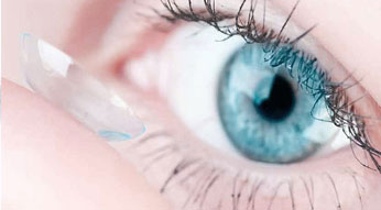
Rigid (hard) gas-permeable (RGP) contact lenses can greatly improve vision when glasses are no longer effective.

These are the most common method of improving the vision for patients with Keratoconus. Supported by the natural tears in the eye, the irregular surface of the cornea is reshaped allowing for better vision.

The development of special keratoconus RGP contact lens designs has increased the usage of RGP lenses over the years. However, RGP contact lenses cannot be worn by everyone. Many people will experience unacceptable discomfort. Therefore they are only partially successful, especially for patients with low production of tears, as an adequate supply of tears is needed to provide adequate lubrication to buffer the RGP lens.

Piggyback or Hybrid lenses

If RGP contact lenses are not tolerated, piggyback lenses are sometimes used, where the hard contact lens is placed on top of a soft contact lens making wear more comfortable.

This superimposed method, although more awkward, may sometimes be better tolerated than an RGP contact lens alone.

Another option is the use of a hybrid lens (hard centre and soft edge), which can be an alternative solution.

Scleral lenses

In very advanced cases of Keratoconus, a well fitted scleral contact lenses may offer visual correction.

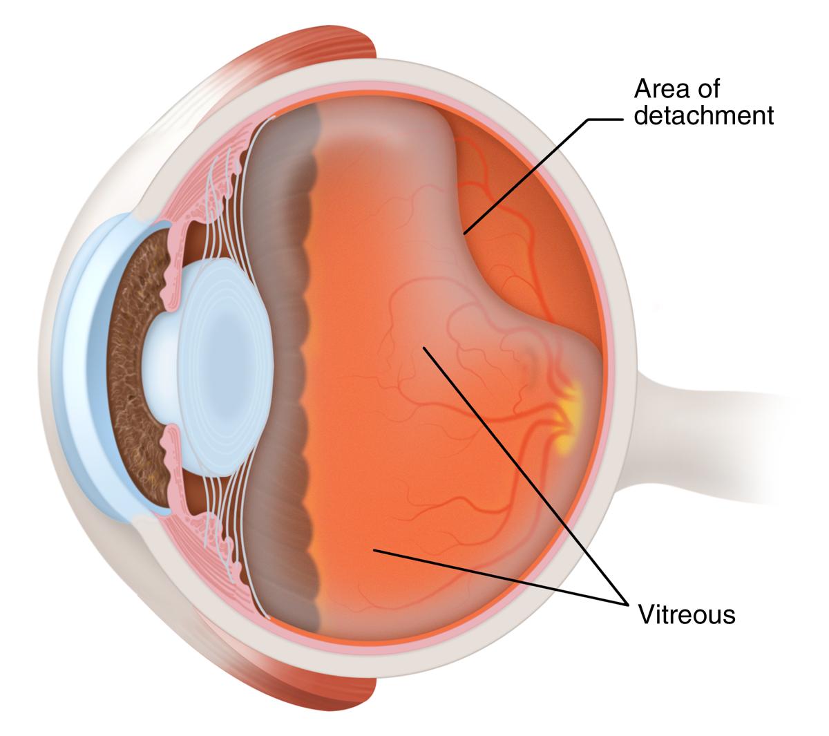
The retina is the lining of the back of the eye, which allows the eye to see. If a hole appears in the retina it will detach rather like wallpaper peeling off a wall. The retina cannot work when it is detached. The only way to repair the retina is by operation to find the hole or holes and sealing them.

Certain symptoms may indicate retinal disease, these include loss of vision, visual distortion, flashes and floaters, a shadow in the vision, blurred vision and / or patchy vision. Patients experiencing these symptoms should seek advice from an opthlamologist.

Treatment

The retina can be repaired in one of two ways:

  1. The hole can be sealed by sewing a small piece of plastic onto the outside of the eye, creating a dent in the eye ball, which will close the hole. You may be able to feel the plastic on the eye after the operation.
  2. Alternatively, it is possible to go inside the eye and, by removing the jelly in the eye (known as the vitreous), a gas bubble can be inserted to support the retina. This procedure is known as a vitrectomy and is performed by making three small incisions in the eye. The operation takes approximately one hour to perform. The gas bubble will float inside the eye and close the hole. Laser or freezing treatment is used to seal the hole.

The surgeon will decide which type of surgery is most appropriate for each individual patient. The eye does not need the vitreous jelly – it will fill the space with a watery fluid.

Posterior vitreous detachment (PVD) is the most important event in retinal surgery. It causes many of the conditions seen by a retinal surgeon. It is very common over the age of 40 years and more often in myopes. PVD may cause symptoms of floaters and flashes in the eye. If you suffer a sudden onset of these symptoms you should seek examination of your eyes by a retinal specialist. In 10% of patients with acute PVD a retinal tear can be found and will need treatment with laser. Untreated retinal tears can lead to retinal detachment which can severely reduce your vision.

What is macular degeneration?

This is an aging change in the eye which damages the macula of the retina thereby reducing central vision. Unfortunately, macular degeneration is common in the elderly and can cause irreversible central visual loss. Peripheral vision is maintained even when the patient has lost reading vision.

There are two main types:

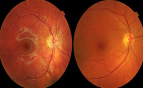
maculardegeneration_clip_image002Dry: Most patients with macular degeneration suffer from the dry form of the degeneration producing a gradual weakening of the retina. This can be seen as yellow deposits in the retina called Drusen or as areas of increased or decreased pigmentation of the retina. It is important to have the eye examined as for some types of dry degeneration (for example the eye on the right) special vitamin and antioxidant tablets have been found to reduce the risk of progression to visual loss.

It is useful to seek advice on the severity of your macular degeneration as the risk of progression to visual loss varies from 3% to 40% over 5 years depending on the appearance of the retina.

maculardegeneration_clip_image006Wet: A few patients will produce a more severe form where a blood vessel membrane appears in the eye as shown on the left. This causes damage to the retina with severe loss of vision often in both eyes. In the early stages the vision distorts followed later by loss of central vision.

Anti VEGF Injections

Anti VEGF drugs, namely Ranibizumab (Lucentis) and Aflibercept (Eylea), restrict the growth of blood vessel membranes in wet ARMD. These block the action of a hormone in the retina called VEGF which makes blood vessels grow. The drugs are injected into the eye under local anaesthesia and have been extremely useful for maintaining or improving vision in this otherwise blinding condition.
The use of these drugs may involve monthly injections and repeat injections over years.

What is the Macula?

The macula is the part of the retina which is needed for fine detail such as reading or recognising faces. It contains delicate receptors called cones which also provide colour vision. Distortion of vision (straight lines appearing to look bent, check your eyes by observing a rectangular window frame one eye at a time) is a common symptom of macular disease.

Macular Hole

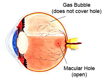
A hole can appear in the macula. This has a tendency to distort and reduce the vision. It is possible to close the hole to improve the vision by vitrectomy operation. This involves removing the jelly of the eye (vitrectomy) and inserting a gas bubble which remains in the eye for a few weeks. The gas bubble floats in the eye and pushes on the hole.

Please note that extreme posturing regimes (e.g. face down) can be avoided with modern macular hole surgery, and is rarely used by Professor Williamson.

  • There is a chance the eye will develop a cataract in the next few years.
  • The patient must not travel by plane whilst the gas bubble is in the eye.
  • 2-6 months may be required for the eye to recover its best vision after surgery.

Macular Pucker

Sometimes a membrane appears that wrinkles the retina – this is known by different names including cellophane maculopathy, epiretinal membrane and macular pucker. A macular pucker blurs your vision and causes distortion (bending or twisting of straight lines). It is possible to operate to try to remove the membrane. The operation involves removing the jelly of the eye (by way of a vitrectomy) and surgically peeling the membrane off the retina. This operation is usually effective in reducing the distortion and improving the vision. A cataract often appears afterwards.

Diabetic retinopathy is a condition whereby damage occurs to the small blood vessels in the retina of the eye. This may cause swelling in the retina, ischaemia (low oxygen) in the retina and more severe complications, which can lead to blindness.

The mainstays of treatments are laser therapy to the retina or injections of Anti-VEGF drugs into the eye. In some patients with diabetes more extensive surgery is required – there are two main reasons why a patient with diabetes may need surgery:

  • Diabetic Haemorrhage; and / or
  • Diabetic Retinal detachment

Diabetic Haemorrhage

Diabetes causes abnormal blood vessels to appear in the eye. Laser treatment is used to try to prevent these occurring. These blood vessels are fragile and bleed easily. Sometimes they bleed into the jelly of the eye (the vitreous) reducing vision. Where this has occurred, the vitreous and the bleed are removed by way of an operation known as a vitrectomy and more laser is subsequently applied.

Diabetic Retinal Detachment

Abnormal blood vessels produce scarring which in turn can produce a retinal detachment. To treat this the vitreous is removed and the scars are dissected off the retina. A bubble of gas may be used to support the retina.

When operating on an eye with diabetic retinal detachment, there is a chance a cataract will develop within the next few years.

Post operatively, these patients are prohibited from travelling by plane whilst a gas bubble is in the eye. Your surgeon will advise you when it is safe for you to fly again.

The eye may suffer inflammation or infection requiring intervention by the vitreoretinal surgeon.

What is inflammation?

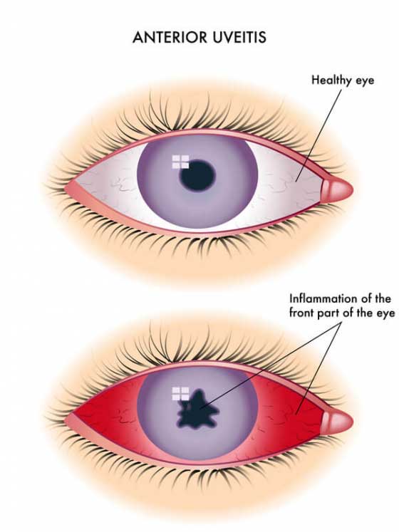
Sometimes the body may produce a reaction against itself. This happens commonly in the joints or skin and also in the eye. This produces a condition called uveitis.
Uveitis usually only affects the front of the eye and can be treated by drops. Sometimes uveitis affects the back of the eye including the retina and vitreous. When this happens the retina can be damaged and the vitreous become opaque with proteins and cells. These may require vitrectomy surgery to repair.
What kind of infections affect the retina?
Almost all kinds of organism can infect the back of the eye:

  • Bacteria after trauma or surgery
  • Viruses giving retinal blood vessel inflammation
  • Fungi such as candida
  • Others such as toxoplasma and toxocara

The retinal surgeon may be required to insert antibiotics or to remove the infection by vitrectomy.

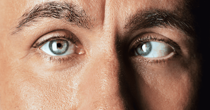
To understand how strabismus surgery works, consider that each of your eyes has six outside (extraocular) muscles controlling eye movements.

If a muscle is too strong when you have strabismus, it may cause the eye to turn in, turn out or rotate too high or low.

On the other hand, an eye muscle weakness in certain cases may also cause misalignment. This condition may occur if you have a

cranial nerve

 dysfunction affecting how eye muscles control movement.

Fortunately, your ophthalmologist has various surgical options to help correct these types of problems.

Strabismus Surgery Involving Recession And Resection Procedures

In a recession procedure, your eye surgeon detaches the affected outside muscle (extraocular muscle) from the eye and reattaches it (resection) farther back on the eye to weaken the relative strength of the muscle if it is too strong.

muscles of the eye

Muscles of the eye.

In contrast, if the muscle is too weak, your surgeon may use a recession procedure to reduce strength of the opposing muscle (antagonist) to achieve more balanced function of the eye muscles.

In certain cases, a resection procedure may be used to strengthen an eye muscle to correct misalignment associated with strabismus. If you have inwardly turned eyes (

esotropia

), the surgeon may strengthen the lateral rectus muscles — located on the side of each eye, toward the ear — by reattaching the muscle in a different location (resection). In this way, the lateral rectus muscles are relatively strengthened and they can turn the eyes farther outward. This results in better eye alignment.

Adjustable Suture Strabismus Surgery

With adjustable suture eye muscle surgery, your surgeon adjusts sutures holding eye muscles in place after a resection procedure, to attempt to improve your final outcome.

Generally this surgery is possible only in adults, with perhaps only a small percentage able to benefit. This surgery is probably best for someone in whom strabismus developed in adulthood after previously normal eye alignment.

In this case, the person is a good candidate because of fusion potential — the ability of both eyes to “lock on” to a target simultaneously, resulting in stereovision and a high degree of depth perception.

In most cases, adjustable suture surgery is performed in the operating room, with general or local anesthesia. Afterward the eye is patched. About four to 24 hours later, the patch is removed in the office, when anesthesia and sedation have faded. Ocular alignment is then evaluated.

Based on how your eyes are aligned, your surgeon may decide to use the suture that is in place to tighten or loosen the treated muscle. This adjustment may cause slight discomfort, primarily with muscle tightening.

Once the desired alignment is achieved, the surgeon ties the adjustable suture permanently in place, and the procedure is complete.

WHAT IS GLAUCOMA?

The optic nerve carries images from the retina (light-sensitive layer at the back of your eye) to your brain, allowing you to see.

Glaucoma is the name given to a group of conditions that cause damage to the optic nerve where it leaves your eye. Glaucoma can cause loss of vision.

Glaucoma can be caused by an increase in pressure in your eye

Sometimes the optic nerve can be damaged, even though the pressure in your eye is within the normal range.

WHAT ARE THE BENEFITS OF SURGERY?

The aim is to reduce the risk of further damage to the optic nerve.

ARE THERE ANY ALTERNATIVES TO SURGERY?

You can use eye drops to lower the pressure.

Laser treatment may be suitable for you but is often less effective than surgery.

WHAT DOES THE OPERATION INVOLVE?

Various anaesthetic techniques are possible, including a general anaesthetic or a local anaesthetic that is injected around your eye to numb it.

The operation usually takes 45 to 75 minutes.

Your surgeon will make a small draining hole in the lining of the white of your eye. The fluid will drain out into a space in the outer lining of your eye and collect just under your eyelid.

WHAT COMPLICATIONS CAN HAPPEN?

GENERAL COMPLICATIONS OF ANY OPERATION

  • Pain
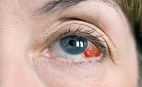
  • Bleeding
  • Infection

SPECIFIC EARLY COMPLICATIONS

  • Heavy bleeding inside your eye
  • Bleeding at the front of your eye
  • Inflammation in your other eye
  • Too much fluid draining
  • Sharp rise in eye pressure

SPECIFIC LATE COMPLICATIONS

  • Developing a cataract
  • Reduced vision over time
  • Failure of the operation

HOW SOON WILL I RECOVER?

You should be able to go home after a few hours.

Your surgeon will need to check your eye the day after the operation. They will see you several times in clinic during the first few weeks and may perform minor adjustments.

Most people will need about 2 weeks off work.

Do not swim, lift anything heavy or bend so your head is below your waist until you have checked with your surgeon.

Regular exercise should help you to return to normal activities as soon as possible. Before you start exercising, ask the healthcare team or your GP for advice.

Most people make a good recovery from the operation, with their glaucoma under better control.

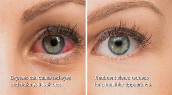
Inability of the eye to produce tears adequately or facing the problem in which the tears produced are not of the correct consistency and quickly evaporates, results in dry eyes.

Inflammation of the eye surface along with dry eye may also occur. Without treatment, it can lead to ulcers, pain, or scars on the cornea, and even loss of some vision. However, a permanent loss of vision due to dry eye is not commonly observed.

It may be difficult to do certain activities such as reading for an extended period of time or using a computer due to this condition. Dry eyes can reduce the tolerance for dry environments, such as the air inside an airplane.

Dry eye condition is known by many names such as dry eye syndrome, keratoconjunctivitis sicca (KCS), lacrimal keratoconjunctivitis, dysfunctional tear syndrome, aqueous tear deficiency, evaporative tear deficiency and LASIK-induced neurotrophic epitheliopathy (LNE).

What are the types of dry eye?

1) Failure of the lacrimal glands to produce adequate amount of the watery component of tears required to maintain a healthy eye surface results in aqueous tear-deficient dry eye.

2) Inflammation of the meibomian glands, located in the eyelids leads to evaporative dry eye. Meibomian glands make up the lipid or oily part of tears and helps slow down the evaporation rate and maintains stability of tears.

Dry eye can be associated with the following conditions:

  • inflammation of eye surface, the conjunctiva, or the lacrimal gland;
  • any disease process that results in alteration of the components of the tears;
  • an increase in the surface of the eye, as in thyroid disease when the eye protrudes forward;
  • cosmetic surgery to correct the wide eyelids.

Causes

Dry eyes can occur due to the following causes:

  • Aging largely contributes to the development of dry eyes. Majority of people above the age of 65 develop dry eye symptoms.
  • Hormonal changes due to pregnancy, menopause and the use of oral contraceptives lead to a greater number of women developing dry eyes as compared to men.
  • Medications such as antihistamines, blood pressure medications, decongestants, and antidepressants, can lead to a reduction in tear production.
  • Having rheumatoid arthritis, thyroid problems diabetes, problems with eyelid inflammation (blepharitis), inflammation of eye surface or the inward or outward turning of eyelids can lead to development of dry eyes.
  •       Wind and dry climates or exposure to smoke can result in an increased evaporation of tear causing dry eye symptoms
  •             Not blinking regularly while working on computer screen for a long time can also result in drying of the eyes.
  •             Long-term use of contact lenses and refractive eye surgeries, such as LASIK, can reduce tear production and contribute to dry eyes.

What is Cosmetic Eye Surgery ?

Medically known as double eyelid surgery or Blepharoplasty, is the type eyelid plastic surgery which is done for the removal of excess fat from the lower eyelids and the upper eyelids. The cause of droopy eyelids is the sagging skin that can also harm the vision and is also the result of excessive accumulation of the fat cells. The aim of the cosmetic surgery is to restore and heal the congenital defects or the disfigurement in order to give a better appearance to the body. Drooping eyes and puffy eyes are the most common cosmetic problems.

Cosmetic Eyelid Surgery

  • Evaluation of eyelid laxity
  • Lower and upper eyelid Blepharoplasty
  • Ptosis repair
  • Management of the dry eye

Facial Cosmetic Surgery Around the Eyelids and Eye

  • Skin Resurfacing
  • Direct Browlift

Orbital Reconstructive Surgery Including Surrounding Bones of the Eyes

  • Grave’ disease (thyroid) surgery
  • Repair and evaluation of lacrimal system
  • Orbital reconstruction by following trauma

Surgical Procedures of Cosmetic Eye Surgery

Brow Lift : The small incisions are made above the hairline in order to improve the appearance of the eyebrow. Also referred to as forehead lift surgery, the aim of the surgery is to raise the eyebrows to offer more youthful and alert look. The surgery also reduces the creases that develop between the eyes, across the forehead and on the bridge of the nose. The sagging brow which is there at the upper eyelid is also repositioned.

Transconjunctival Blepharoplasty Eye Lift : The excessive accumulation of the fat is repositioned or removed by making a small incision in the eye that has caused eyelid bags. The incision made inside the lower eyelid can also be done with resurfacing.

Lower Eyelid Lift Surgery : Blepharoplasty or lower eyelid lift surgery is performed to remove the excess skin, muscle, fat, wrinkles and puffy fat bags of the lower eyelids. The puffy fat bags on the lower eyelid makes us look more sad, tired and old. The aim of the surgery is to correct this by making an incision below the eyelid. This surgery gives the patients more alert, rested and youthful appearance.

Upper Eyelid Lift Surgery : During the surgery, incisions are made along the natural fold of the upper eyelid that widens beyond the existing creases or into the laugh lines. With the help of incisions, the excess fat, muscle and skin is removed. As the incisions are done on the natural contour of the upper eyelid, they are not noticeable.

Resurfacing Surgery : This surgery is primarily done to treat eye wrinkles.

Laser Eyelid Surgery : This surgery involves the use of CO2 (carbon dioxide) laser in order to remove the excess fatty tissue and skin. Small incisions are made with the help of the ultra pulse laser that instantly ablates with the tissues surrounding the lower and upper eyelids. The aim of the surgery is to tighten the saggy and loose skin by eliminating the bags under the eye.

Canthoplasty : This particular type of surgery is performed to tighten the lower eyelid of the patient. It comes under Blepharoplasty, which is a surgical procedure done to modify the eyelids. The surgical procedure is also known as inferior retinacular suspension, it is used to alter the eye shape permanently. Canthoplasty is the most popular procedure for those who want more youthful shape of the eyes. The surgery is also meant for those who suffer from trauma or droopy eyelids. Canthoplasty can be simultaneously done with eyebrow lift or face lift procedures.

Make An Appointment With Us

    Our Doctors

    The specialists at Alnoor Eye Centre have undergone advanced training and have years of experience treating cataracts, refractive errors, corneal disease, retinal disease, glaucoma, eye cancers and other eye conditions.

    Prof. Dr. Samina Jahangir

    MBBS (Gold medalist) FCPS (Gold medalist) MD (USA)

    Prof. Dr. Tehmina Jahangir

    MBBS (KEMC) MRCOphth (UK) MCPS, FCPS (Ophthalmology)

    Associate Prof. Dr. Sana Jahangir

    MBBS (Aga Khan University) MD (USA) FCPS (Ophthalmology) FCPS (Vitreoretinal Surgery)

    Call Now Button
    WhatsApp chat